Cataract Surgery
A cataract is a clouding of the eye’s natural lens, which lies behind the iris and the pupil. Cataracts are the leading cause of blindness worldwide, responsible for approximately 51% of global blindness, according to the World Health Organization. As the population ages, the prevalence of cataracts is expected to rise, making it a significant public health concern.
Cataracts can severely impact quality of life, leading to visual impairment, loss of independence, and increased risk of falls and injuries. However, cataract surgery is one of the most common and successful procedures performed worldwide, with high rates of improved vision. This paper aims to provide a detailed overview of cataracts, including their types, pathophysiology, clinical presentation, and management strategies.
Cataracts can be classified based on their location within the lens, their cause, or the age at which they develop. The main types of cataracts include:
- Nuclear Sclerotic Cataract:
- This type of cataract forms in the nucleus, the central part of the lens. It is the most common type of age-related cataract and is characterized by the gradual hardening and yellowing of the lens nucleus. Patients with nuclear sclerotic cataracts often notice a decrease in distance vision and an increase in near vision, a phenomenon known as “second sight.” As the cataract progresses, it can lead to significant visual impairment.
- Cortical Cataract:
- Cortical cataracts develop in the lens cortex, the peripheral part of the lens, and are characterized by the presence of white, wedge-shaped opacities or “spokes” that radiate from the periphery toward the center of the lens. These opacities scatter light, causing glare and difficulty with night vision. Cortical cataracts can cause fluctuating vision as the opacities progress.
- Posterior Subcapsular Cataract:
- This type of cataract forms just in front of the posterior capsule, the back surface of the lens. It is often associated with steroid use, diabetes, and radiation exposure. Posterior subcapsular cataracts can cause significant visual impairment, particularly in bright light, and are often associated with glare and difficulty reading.
- Congenital Cataract:
- Congenital cataracts are present at birth or develop during infancy. They can be caused by genetic factors, intrauterine infections (such as rubella), or metabolic disorders. Congenital cataracts can affect one or both eyes and, if left untreated, can lead to amblyopia (lazy eye) and permanent vision loss.
- Traumatic and Secondary Cataracts:
- Traumatic cataracts result from direct injury to the eye, such as blunt or penetrating trauma, electrical injuries, or exposure to radiation. Secondary cataracts, also known as “after-cataracts,” can develop after cataract surgery, particularly in younger patients, due to the proliferation of lens epithelial cells on the posterior capsule. Secondary cataracts can also result from systemic diseases like diabetes or prolonged use of medications such as corticosteroids.
Cataract formation is a multifactorial process involving complex biochemical and structural changes within the lens. Several factors contribute to the development of cataracts:
- Risk Factors: Age is the most significant risk factor for cataract development, with most cases occurring in individuals over 60. Other risk factors include ultraviolet (UV) radiation exposure, smoking, diabetes, hypertension, and prolonged use of corticosteroids. Lifestyle factors, such as poor diet and excessive alcohol consumption, can also contribute to cataract formation.
Cataracts typically develop slowly and may initially be asymptomatic. However, as the cataract progresses, it can lead to a variety of symptoms that affect the patient’s quality of life:
- Symptoms:
- Patients with cataracts often report blurred or dim vision, which cannot be corrected with glasses or contact lenses. They may also experience increased sensitivity to light and glare, particularly when driving at night. Other common symptoms include seeing halos around lights, frequent changes in prescription glasses, double vision in one eye, and fading or yellowing of colors. As the cataract progresses, the patient’s visual acuity continues to decline, making everyday tasks increasingly difficult.
- Signs:
- During a slit-lamp examination, cataracts present as opacities within the lens. The appearance of the cataract varies depending on its type In advanced cases, the entire lens may become opaque.
- Diagnostic Approaches:
- Visual Acuity Testing: The standard initial test to assess the impact of the cataract on the patient’s vision. It involves reading an eye chart to determine the clarity of vision at various distances.
- Slit-Lamp Examination: A slit-lamp biomicroscope provides a magnified view of the eye structures, allowing the ophthalmologist to examine the lens for cataracts and assess their type, size, and location.
- Dilated Eye Exam: Dilating the pupil with eye drops enables a more thorough examination of the lens and the back of the eye, helping to identify any additional eye conditions that could affect the choice of treatment.
- Diagnostic Imaging: Optical coherence tomography (OCT) and B-scan ultrasonography may be used in certain cases to assess the lens’s condition and other ocular structures, particularly if there is a dense cataract or coexisting retinal pathology.
The management of cataracts depends on the severity of the symptoms and the impact on the patient’s quality of life. Treatment options range from conservative management to surgical intervention:
- Non-Surgical Management:
- Use of Glasses and Magnifying Aids: In the early stages of cataract formation, adjusting the patient’s eyeglass prescription can help improve vision. Magnifying lenses, stronger lighting, and anti-glare sunglasses can also alleviate some of the visual disturbances caused by cataracts.
- Lifestyle Modifications and Patient Education: Patients should be advised to avoid smoking, reduce alcohol consumption, and protect their eyes from excessive UV exposure by wearing sunglasses. Maintaining a healthy diet rich in antioxidants, such as vitamins C and E, can also help slow the progression of cataracts.
- Surgical Management:
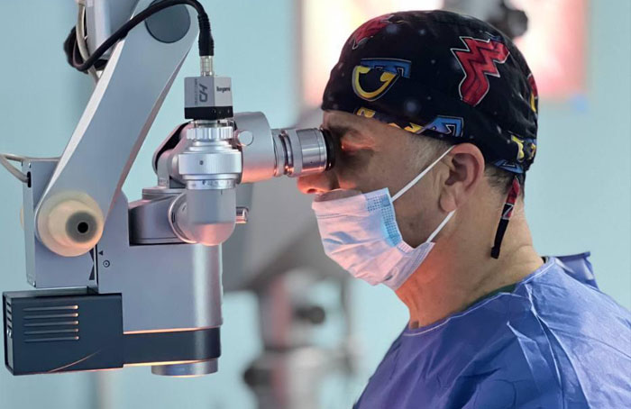
- Phacoemulsification and Intraocular Lens Implantation: Cataract surgery is the most effective treatment for cataracts and involves removing the clouded lens and replacing it with an artificial intraocular lens (IOL). Phacoemulsification is the most common technique, where an ultrasonic device is used to break up the lens into small fragments, which are then removed through a small incision. The IOL is then implanted to restore vision.
- Types of Intraocular Lenses (IOLs): Types of IOLs:
- Monofocal IOLs: Provide clear vision at one distance, typically set for distance vision, requiring patients to use reading glasses for near tasks.
- Extended Depth of Focus (EDOF) intraocular lenses (IOLs) are an advanced type of artificial lens used in cataract surgery and refractive lens exchange. These lenses are designed to provide clear vision at both distant and intermediate ranges, reducing the need for glasses after surgery.
- Multifocal IOLs: Designed to provide clear vision at multiple distances, reducing or eliminating the need for glasses. These lenses can have rings or zones for different focusing powers.
- Toric IOLs: Specifically designed to correct astigmatism, improving overall visual outcomes for patients with pre-existing corneal astigmatism.
- Advanced Surgical Techniques: Femtosecond laser-assisted cataract surgery (FLACS) is an advanced technique that uses a laser to perform key steps of the surgery, such as creating incisions, opening the lens capsule, and fragmenting the lens. This approach can improve the precision of the surgery and reduce recovery time.
- Postoperative Care and Management of Complications:
- After cataract surgery, patients are typically prescribed antibiotic and anti-inflammatory eye drops to prevent infection and reduce inflammation. Follow-up visits are necessary to monitor the healing process and assess visual acuity. Although cataract surgery is highly successful, complications can occur, such as posterior capsule opacification (PCO), cystoid macular edema, and, rarely, endophthalmitis. Early detection and management of these complications are crucial for optimal visual outcomes.